Time-Lapse Video Reveals How Cardiac Cells Team Up To Build a Heart
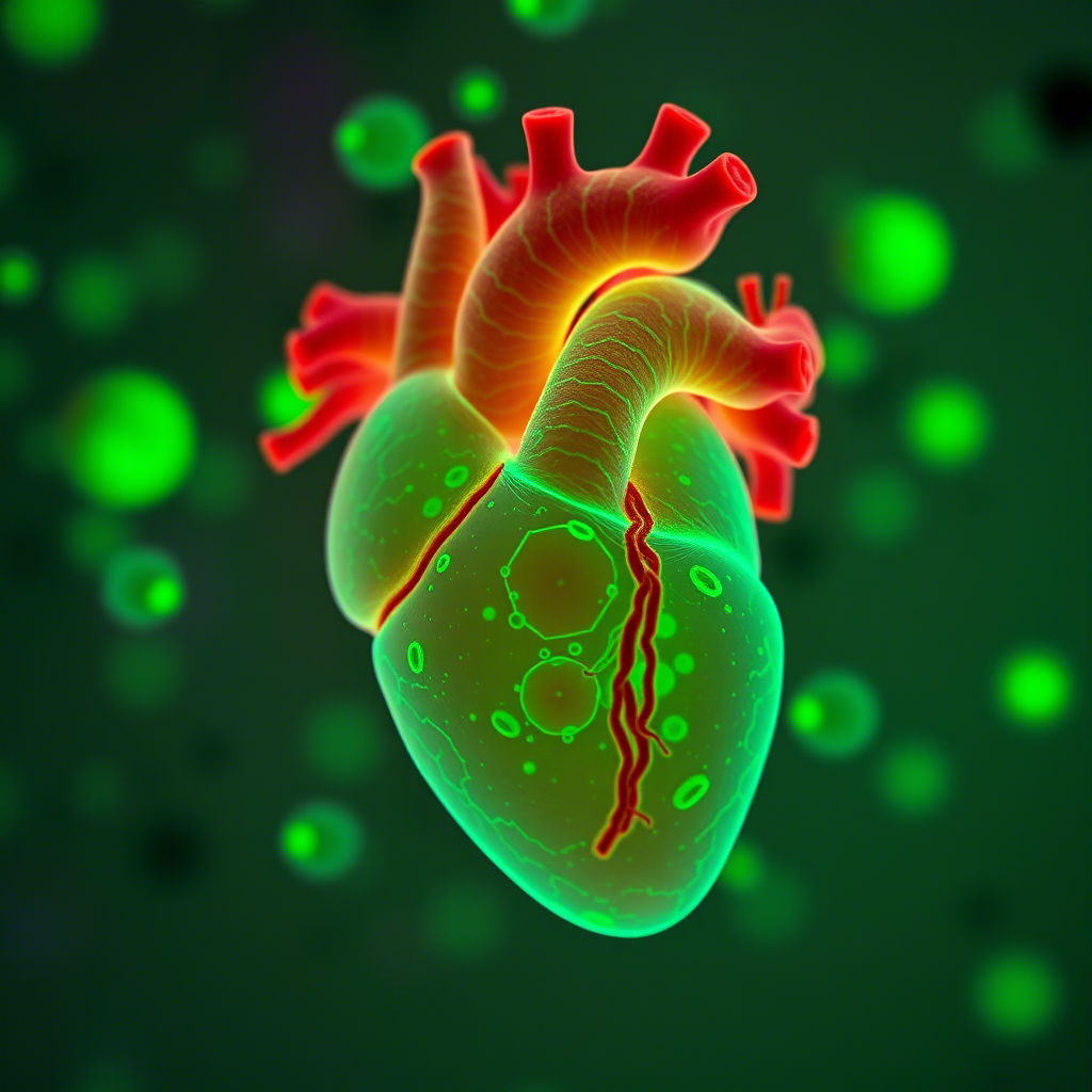
In a stunning new timelapse, scientists have captured the complex process of cardiac cells organizing themselves into a functioning heart during the early development of a mouse embryo.
The footage was made possible using a technique called light-sheet microscopy (LSM), which uses a thin slice of light to scan living tissue, producing high-resolution 3D images without harming the sample.
Researchers from University College London (UCL) and the Francis Crick Institute in the UK used this method to follow how embryonic cells begin to divide, differentiate, and assemble into the early structure of the heart.
By labeling different cell types with fluorescent markers and capturing images every two minutes over 41 hours, the team produced a video showing how a cluster of simple, undifferentiated cells gradually transforms into a fully beating mouse heart — an awe-inspiring glimpse into the origins of life.
This research isn’t just visually stunning — it also revealed surprising insights into how hearts develop. The team found that individual cells seem to “know” their future roles and destinations as early as four to five hours after the first embryonic cell division.
“Our results suggest that the process of determining which cells become heart tissue, and where they end up, starts much earlier than previously thought,” explains Kenzo Ivanovitch, a developmental biologist at UCL.
“This changes how we view heart development — what looked like random cell movement is actually guided by hidden, organized patterns that ensure the heart forms correctly.”
Although practical applications are still far off, this deeper understanding could one day lead to new treatments for congenital heart defects.
The study was published in The EMBO Journal .


